|
|

| What does a Centriole look like? Could you submit
a picture, please? |
| Question Date: 2019-02-13 | | Answer 1:
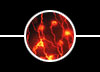
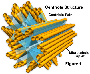
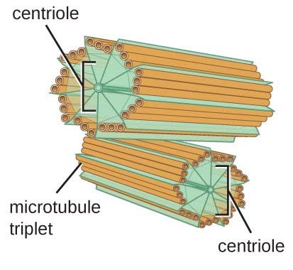
Here is an illustration of a centriole pair:
centriole
1
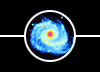
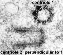
In real life (using a very fancy microscope), they
look very similar:
centriole
2
What you're seeing in this image is the top of one
centriole (lower left) and the side of its partner
centriole (upper right). Each dark circle/rod
is a microtubule. Everything else in the image
is cytoplasm.
Isn't it cool how organized the structure is,
and how we can take pictures of something so
small?!?!
| | Answer 2:
I can't send you a picture, but I could find some
transmission electron micrographs of centrioles
online (see links below). These pictures were
taken by shining an electron beam through the
centriole and having it absorb electrons (using
something called a "transmission electron
microscope"), in much the same way that you
shine X-rays through bones to see what is inside.
This is necessary because centrioles are really
small. I could not find any photographs of
centrioles taken using an ordinary light-using
microscope such as you would use in a classroom,
and this is probably the reason.
Here's the image:
here.
Here's the Northwestern University website from
which the image was taken:
NorthWestern Univ website.
| | Answer 3:
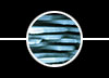
Here's an electron microscope picture of 2
centrioles - the top one is lying on its
side and shows the microtubules lying next
to each other in a circle. The bottom one is
perpendicular to the other, and you can see the
tops of all the microtubules. These microtubules
are in triplets - 3 little o's in a line, and 9 of
the little lines of triplets.
centriole
3
In cell biology a centriole is a cylindrical
organelle composed mainly of a protein called
tubulin. Centrioles are found in most
eukaryotic cells. A bound pair of
centrioles, surrounded by a shapeless mass of
dense material, called the pericentriolar
material, makes up a structure called a
centrosome (from Wikipedia).
| | Answer 4:
A centriole is an organelle in the cell that
helps the cell divide or make copies of
itself. They’re only found in animal cells
and they’re made of proteins called
microtubules. Without the centrioles, the
chromosomes would not be able to move during
mitosis.
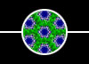
Each centriole is make of nine triplet
groups of microtubules, all grouped together
in a circle. Imagine you have 27 straws. If you
put glued them in groups of threes (in the shape
of a stoplight) and then attached the groups
together in a circle, you’d get a centriole. I’ll
attach a picture below!
image here
Click Here to return to the search form.
|
 |
 |
 |

|
Copyright © 2020 The Regents of the University of California,
All Rights Reserved.
UCSB Terms of Use
|
|
|

